Facilities
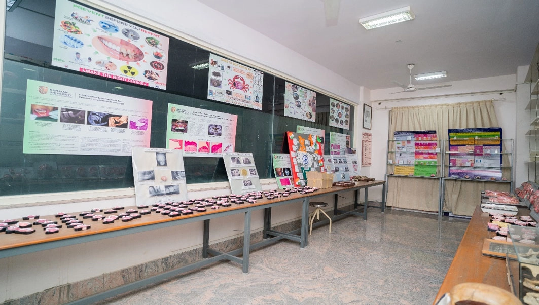
Oral Pathology Museum
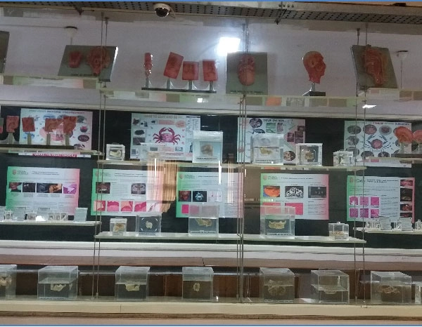
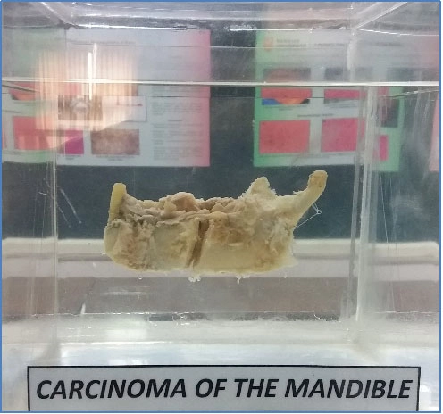
Museum technologies provide a wide array of choice of museums to those who wish to exploit technology to attract, excite and ensure an unrivalled visitor experience, as well as capture and sustain share of mind and heart. Museum being a combination of both art and science requires skilled workmanship, meticulous planning and execution to exhibit a specimen to its optimal elegance due to its relatively smaller size and fragile nature.
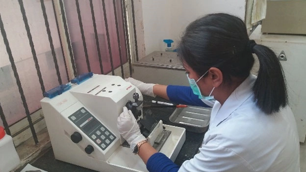
Histopathology
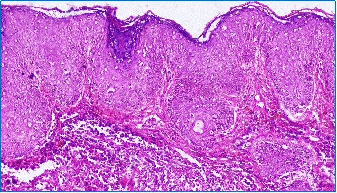
Histology and Histopathology are frequently deliberated and explained together. In fact, the notion of histopathology is inseparable from that of histology. Basic knowledge is important in understanding of normal histology and histopathological interpretation for the diagnosis of lesions.
We provide various histopathological facilities in the department.
Complete provisions for routine histopathological tissue processing, staining and interpretation. Consultation and advice for all histology and pathology enquiries.
Cytology
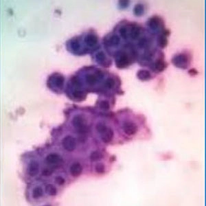
Exfoliative cytology and Fine needle aspiration cytology deals with the microscopic study of cells shed or obtained from the oral mucosa and body fluids for diagnostic purposes. It is a simple, non-aggressive technique that is well accepted by the patient, and is therefore an attractive option for the screening and early diagnosis of infections, benign and malignant lesions.
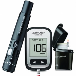
Haematology
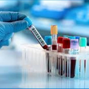
Hematology is the study of blood and blood-forming organs, including the diagnosis, treatment, and prevention of diseases of the blood, bone marrow, and immunologic, hemostatic, and vascular systems. Hematologic analysis is often used prior to invasive dental procedures for the diagnosis and treatment of blood disorders.
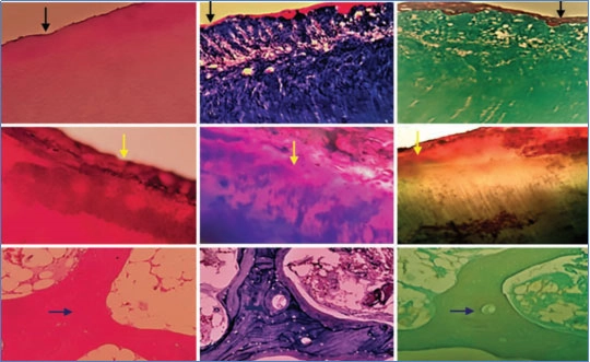
Special Staining
Special stains use a variety of dyes and techniques to stain particular tissues, structures or pathogens to assist pathologists with tissue-based diagnosis.
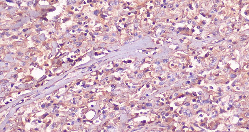
Immunohistochemistry
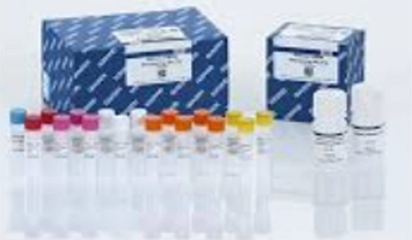
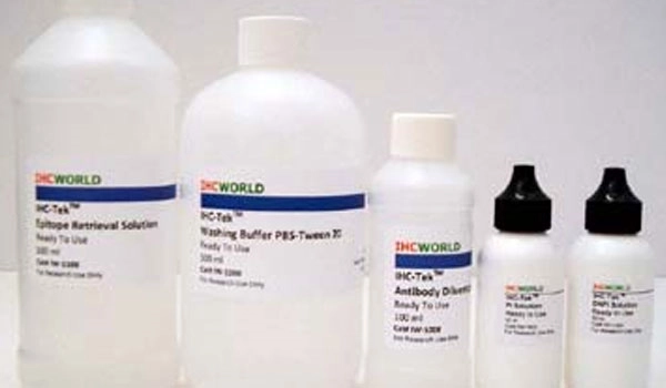
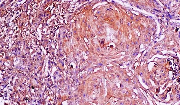
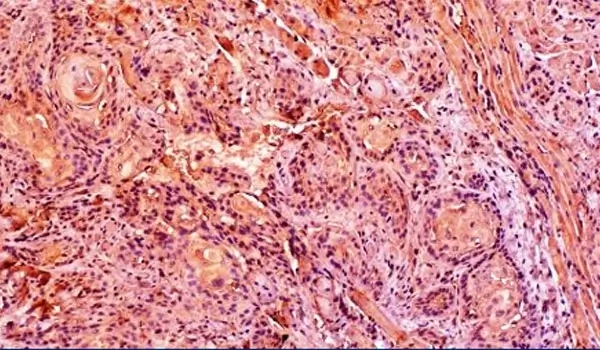
An immunohistochemistry (IHC) lab is a specialized laboratory dedicated to conducting experiments and analyses related to immunohistochemistry techniques. Immunohistochemistry is a powerful method used to detect specific proteins or antigens within tissue sections or cell samples using antibodies and staining processes. It is widely used in various fields of research, including biology, medicine, pathology, and oncology.
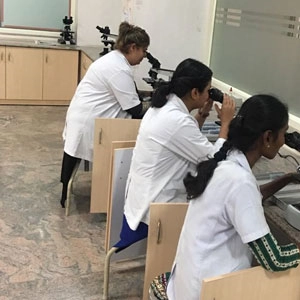
Advanced Microscopy
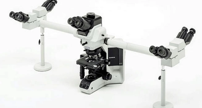
Bright Field Microscope - Penta head attachment(Olympus Optical Microscope BX53F2, Tokyo, Japan)
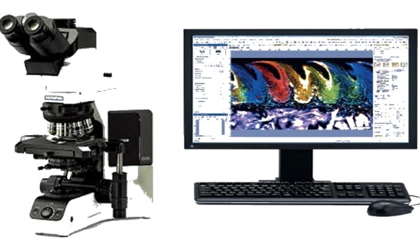
High Resolution Imaging
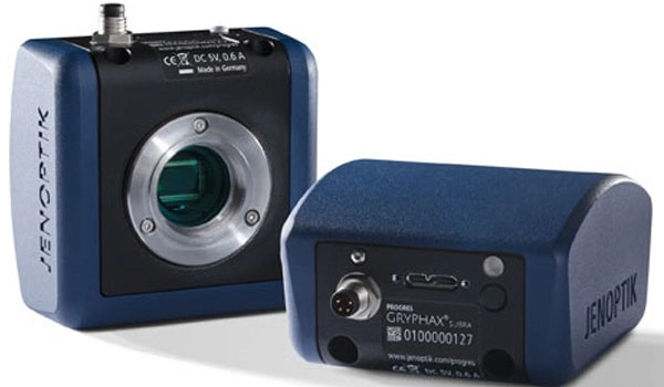
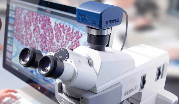
Jenoptik Progres Gryphax Arktur USB 3.0 microscope camera, Jena, Germany
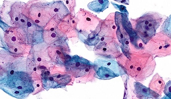
Cytology Imaging
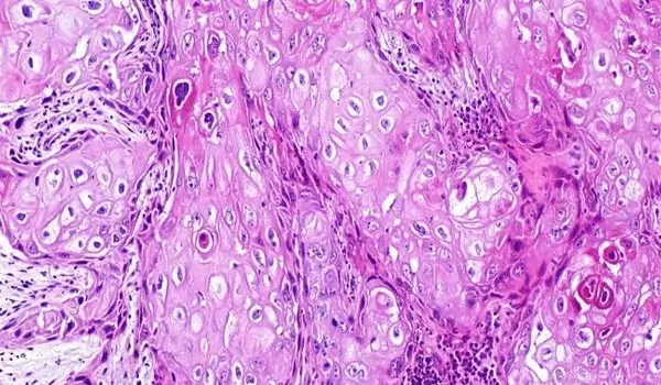
Histopathology Imaging
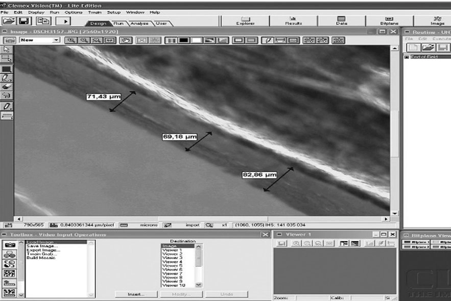
Image Analysis Software
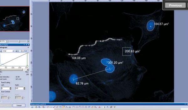
Morphometric Analysis
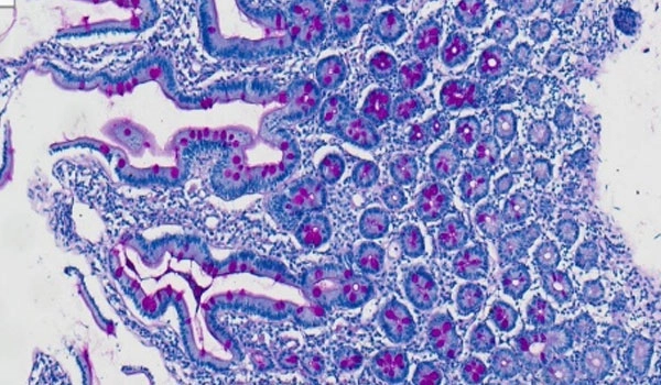
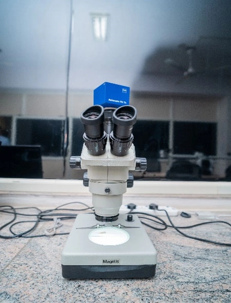
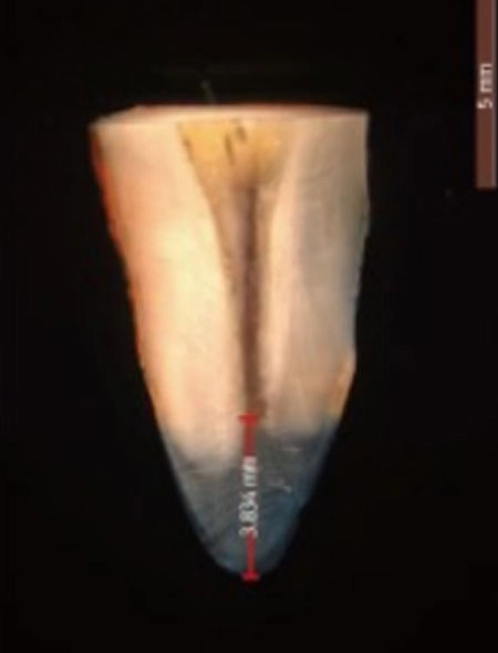
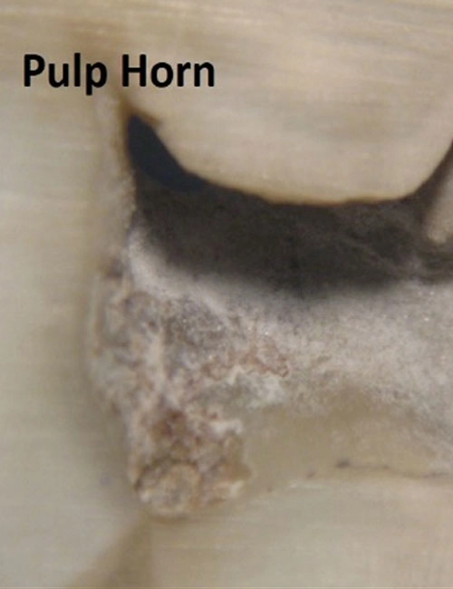
Stereomicroscope
3D Imaging and Morphometry
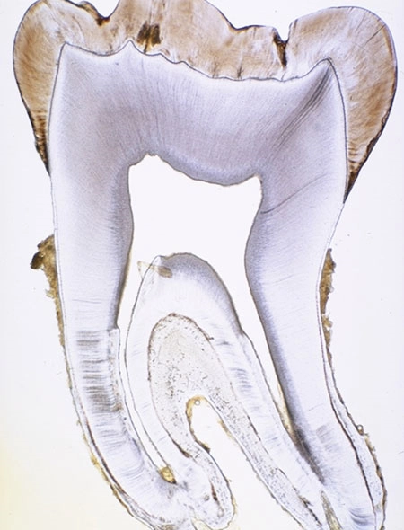
Polarizing Microscopy
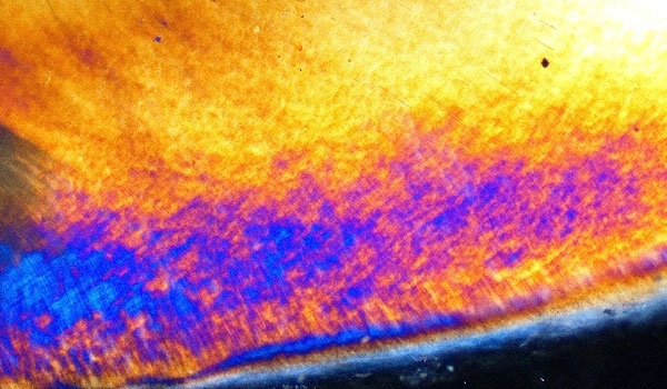
Hard Tissue Studies
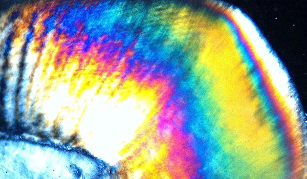
Ground Section of Teeth
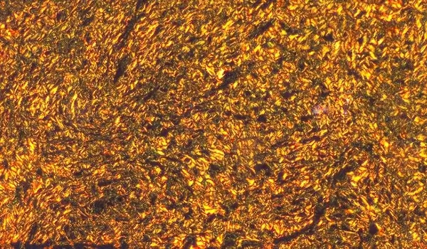
Soft Tissue Studies
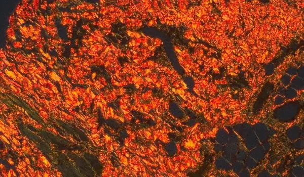
Stained collagen
Phase Contrast and Darkfield Microscopy
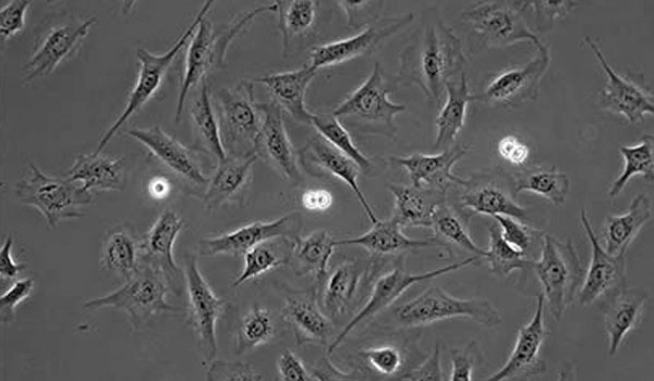
Fibroblasts under Phase Contrast
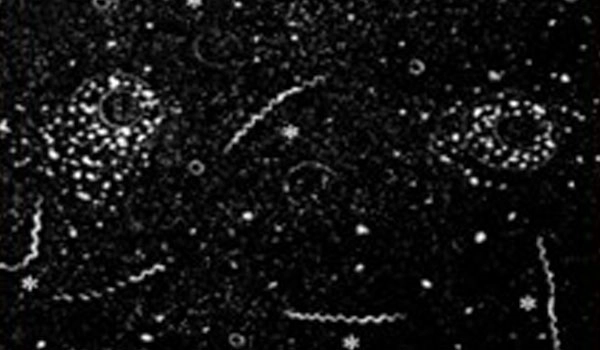
Microorganisms under Dark-Field
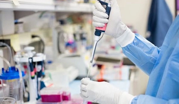
Immunohistochemistry Laboratory
The Immunohistochemistry laboratory offers a variety of services for antigen detection in tissues in the field of surgical pathology and research projects.
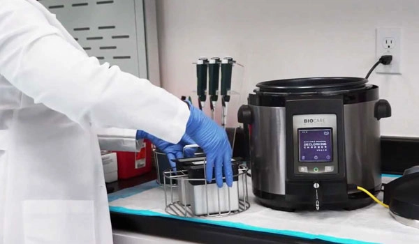
Haematology Laboratory
- Fasting/Random/Postprandial Blood Sugar
- Hemoglobin Estimation
- Differential WBC Count
- Absolute WBC Count
- RBC Count
- Platelet Count
- Erythrocyte Sedimentation Rate
- Bleeding/Clotting Time
- Blood Grouping
- PRF Preparation
- Storage of Samples at – 80C
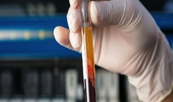
Routine Haematology
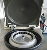
Centrifuge

- 80°C Refrigerator

37°C Incubator
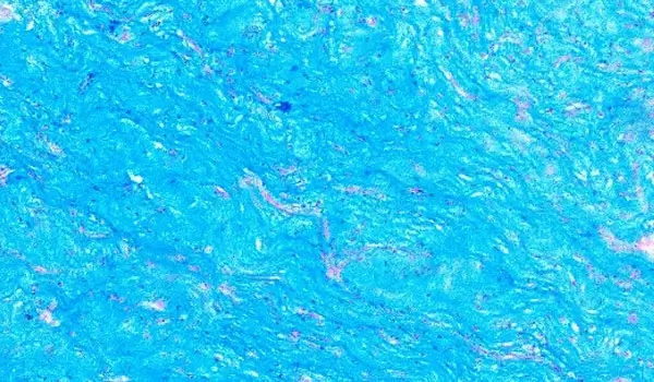
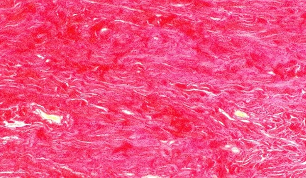
Special Staining of Hard & Soft Tissues
- Periodic Acid Schiff (PAS)
- PAP Stain
- Masson’s Trichome
- Van Gieson
- Alcian Blue
- Picrosirius Red
- Giemsa
- Toluidine blue
- Gram Stain
- Safranin O
- KOH
- Mallory’s
- Ziehl–Neelsen
- Mucicarmine stain
Core Areas of Research
Undergraduate Research
- Analysis of FNAC smears
- Cytological analysis in Oral Cancer
- Chair side smear preparation for fungal infections
- Forensic Odontology
- Assessment of Mitotic figures in Cancer
- Research involving ground section and Decalcified sections
- Special staining to arrive at final diagnosis
- Histotechniques to determine prognosis
- Usage of Specialized Microscopy for pilot studies
- Analysis of Hematological Investigations
Postgraduate Research
- Oral cancer biomarker studies
- Molecular studies in Oral Cancer Tissues
- Histotechniques in Oral Cancer & Pre-cancer
- Clinicopathologic Correlation in Oral Cancer
- Survival Analysis in Oral Cancer
- Analysis of Metastasis in Oral Cancer
- Biofriendly laboratory substitutes
- Biologic behaviour of Odontogenic Tumors and Cysts
- Differential Diagnosis of Salivary gland tumors, developmental disturbances, mesenchymal tumors, skin diseases, bone diseases and infections
Sponsored Research
- Genomic analysis of Oral Cancer & Pre-cancer Tissues
- Artificial Intelligence and Machine Learning
- Photomicrography and Image analysis/processing
- Biomarkers in Oral cancer and pre-cancer
- Biologic Behaviour of Oral Cancer and pre-cancer
- Publications in Impact factor, PubMed and Scopus indexed Journals
